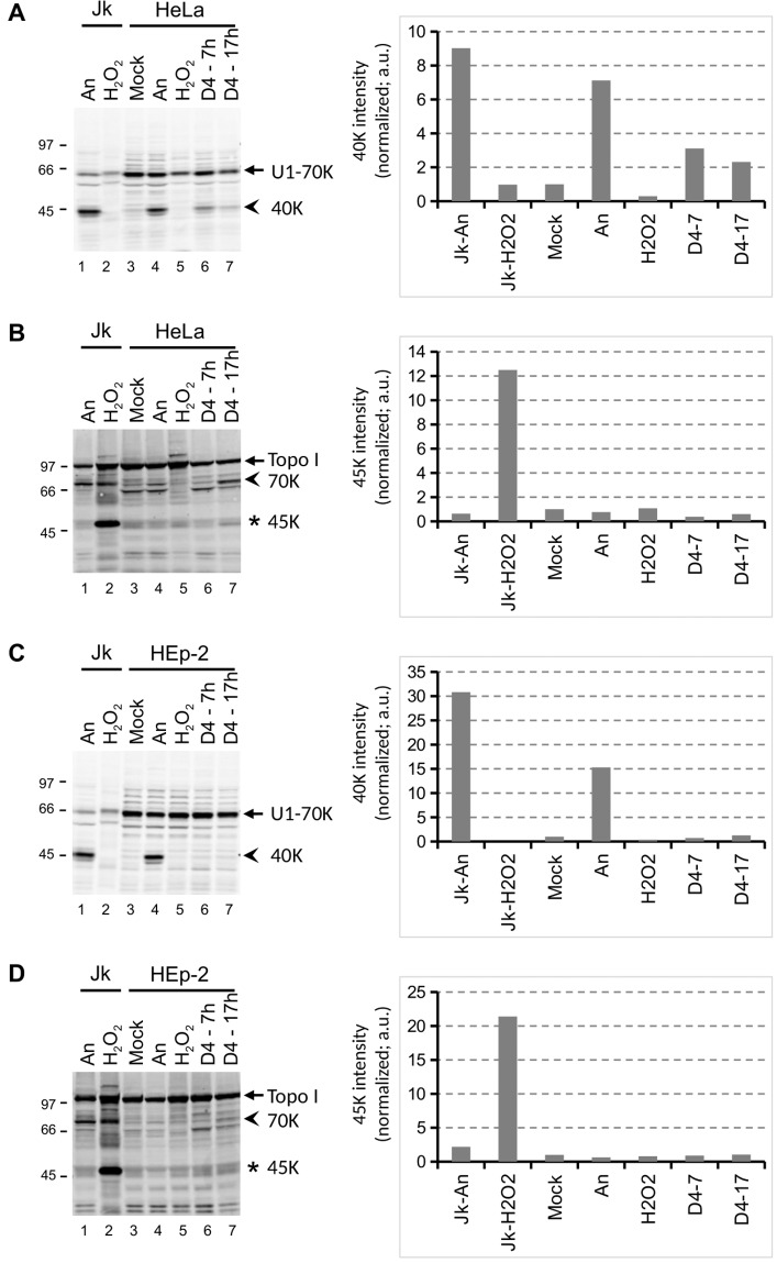
Figure 8.
Cell death induction by D4 in HeLa and HEp-2 cell lines. HeLa (A, B) and HEp-2 (C, D) cells were cultured for the indicated time periods in the presence of 1% D4, or for 4 hours in the presence of 10 μg/ml anisomycin (An) or 0.15% H2O2. Cell lysates were analyzed by western blotting using patient sera reactive with U1-70K (A,C) or Topo I (B,D). The apoptotic cleavage products of U1-70K and Topo I (arrows) are indicated with arrowheads; the necrotic cleavage product of Topo I is indicated with an asterisk. As a reference, material from anisomycin- and H2O2-treated Jurkat cells (Jk) was analyzed in parallel (lanes 1-2). The positions of molecular weight markers are indicated on the left of each panel. Note that in all panels a cropped part of the respective blot is shown, as further illustrated in the Supplementary Information.

