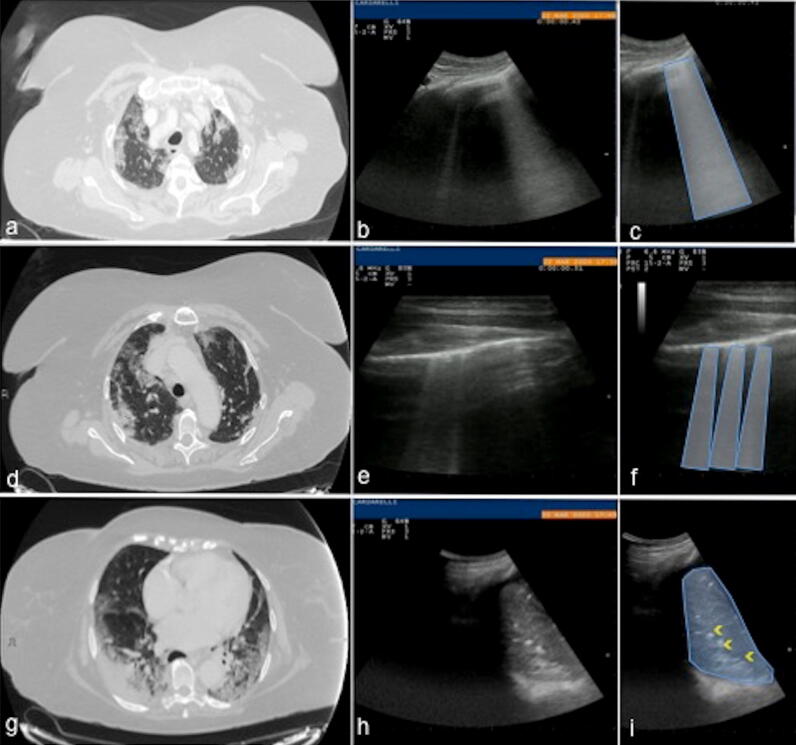
Fig. 15.
Axial chest CT scan in lung window view (a, d, g) at the pulmonary apices (a), upper lobes (d) and lower lobes (g) of a 65 years-old female patient with fever shows: multiple, extensive areas of ground glass opacities in both lung (a, d), coexisting with consolidative opacities and “dark bronchogram sign” (g), consistent with COVID-19 rapid progression stage (positive for COVID-19 nasal-pharyngeal swap RT-PCR). The corresponding ultrasound scans with low-frequency convex probe at right apex (b), linear high-frequency probe at right upper chest zone (e) and convex low-frequency probe at the right lower chest zone (h) show: vertical artifacts originate from the pleura line with a coalescence aspect similar to “white lung” pattern (blue trapezoid, c) at the right apex (b); multiple B lines (f blue trapezoids) at the right superior lobe (e); area of consolidation (i blue area) with air bronchogram (i yellow arrowheads) at the right base (h)

