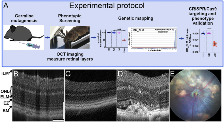
Fig. 1.
Genetic screening protocol and examples of OCT and fundoscopy images from mice with mutations identified in the forward genetic screen. (A) Schematic of the forward genetic retinal phenotype screen including ENU mutagenesis, screening of G3 mice, genetic mapping, and confirmation of gene-phenotype associations by CRISPR/Cas9 targeting. (C/C are mice homozygous for the CRISPR/Cas9 mutation.) (B–D) Representative OCT images for a B6J mouse showing normal retinal structure (B), a mouse with a Cngb1 homozygous mutation showing significant thinning of outer retina (C), and a mouse with a Vldlr homozygous mutation with neovascularization and distorted outer retina (D). (E) Corresponding fundus photo of the same Vldlr homozygous mutant mouse shown in D. BM, Bruch’s membrane; EZ, ellipsoid zone; ELM, external limiting membrane; ONL, outer nuclear layer; ILM, internal limiting membrane.

