Fig. 3.
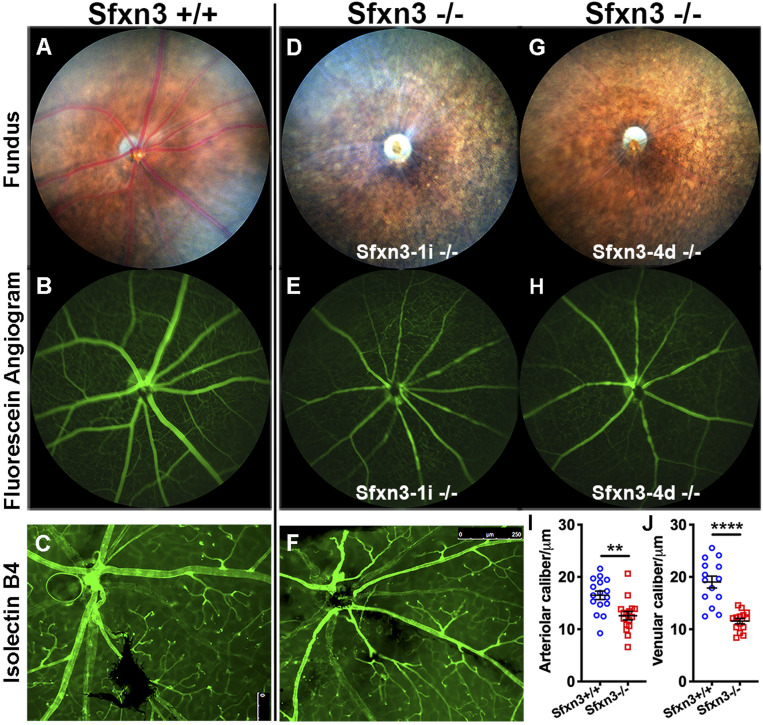
Sfxn3−/− mice show abnormal fundus and vasculature on fundoscopy, fluorescent angiography, and retinal flat mount staining. Sfxn3−/− mice (D–H) show a marked decrease in vessel caliber compared with controls (A–C). The difference is striking on fundus examination (D and G vs. A), in fluorescein angiography (E and H vs. B), and in isolectin B4-stained retinal flat mounts (F vs. C). The difference in caliber is statistically significant when comparing either arterioles (I) or venules (J) measured on the fluorescein angiography images. We did not observe any significant areas of nonperfusion in the Sfxn3−/− eyes. Mice were age 10 to 12 mo (n = 3 mice; 30 vessels/group). All retinal arterioles and venules were measured in three Sfxn3+/+ eyes and three Sfxn3−/− eyes. **P < 0.01, ****P < 0.0001, Student’s t test.

