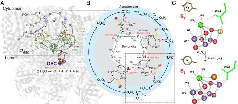
Fig. 1.
The S2 → S3 transition in PS II. (A) The structure of PS II with the membrane-embedded helices and the large membrane extrinsic regions on the luminal side of PS II are shown as cartoon in gray and in color the main electron transfer components involved in charge transfer (P680, Pheo) and stabilization with the acceptor quinones, QA and QB, toward the cytoplasm, and YZ and OEC with catalytic Mn4Ca cluster on the donor side at the interface between the membrane and the lumen-bound part of PS II. (B) Kok cycle of the water oxidation reaction that is triggered by the absorption of photons (nanosecond light flashes, 1F to 4F) in the antenna and the reaction center P680. The white and gray areas show the redox changes of the OEC (S1 to S0) and YZ together with the charge separation reactions, while the outer blue circle shows the concomitant acceptor side chemistry of the quinones. The S2 → S3 transition, marked by the red box, is discussed in detail in the main text. (C) The structures of the Mn4Ca cluster, showing the major changes between the S2 and S3 states (Mn3+ yellow, Mn4+ purple, Ca2+ green, O red; W1, 2, 3, 4 are water ligands to Mn4 and Ca). The Glu189 residue of the D1 protein as a ligand of Ca moves away during the transition with the insertion of oxygen (OX = O2− or OH−) as a new bridging ligand between Mn1 and Ca (adapted from ref. 20).

