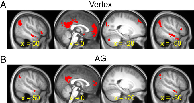
Fig. 5.
fMRI-TMS results: Whole-brain. (A) Shown in red are whole-brain regions demonstrating greater activity for the episodic simulation and divergent thinking tasks relative to the nonepisodic control task following cTBS to the vertex. (B) Whole-brain regions demonstrating greater activity for the episodic simulation and divergent thinking tasks relative to the nonepisodic control task following cTBS to the AG. Results are overlaid on the across-participant mean T1-weighted anatomical image.

