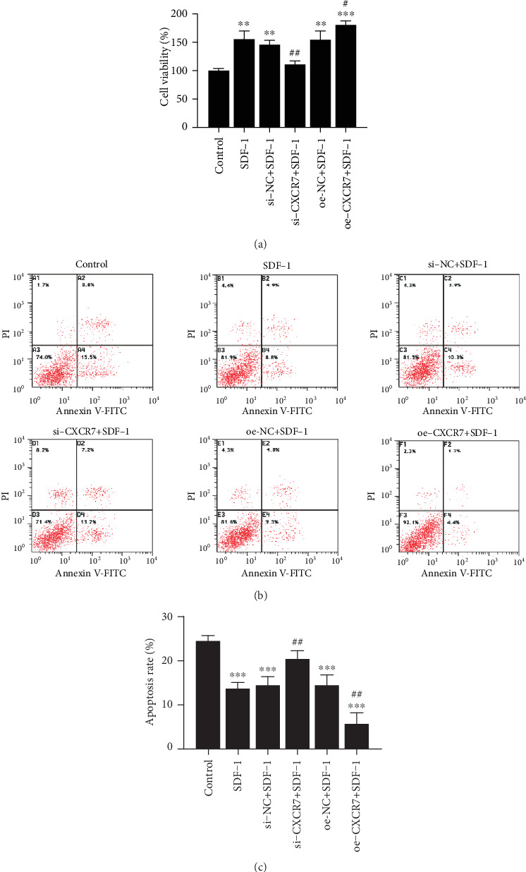
Figure 2.

The effects of CXCR7 on the proliferation and apoptosis of HUVECs. (a) Cells proliferation was measured by CCK-8 at 24 h. (b) HUVEC apoptosis was detected by V-FITC and PI staining. (c) The percentage of apoptotic cells was determined and presented as the mean ± SD. si-NC: siRNA negative control group. oe-NC: overexpression negative control group. ∗∗p < 0.01 versus untreated control group, ∗∗∗p < 0.001 versus untreated control group, #p < 0.05 versus SDF-1 group, ##p < 0.01 versus SDF-1(100 ng/ml) group.
