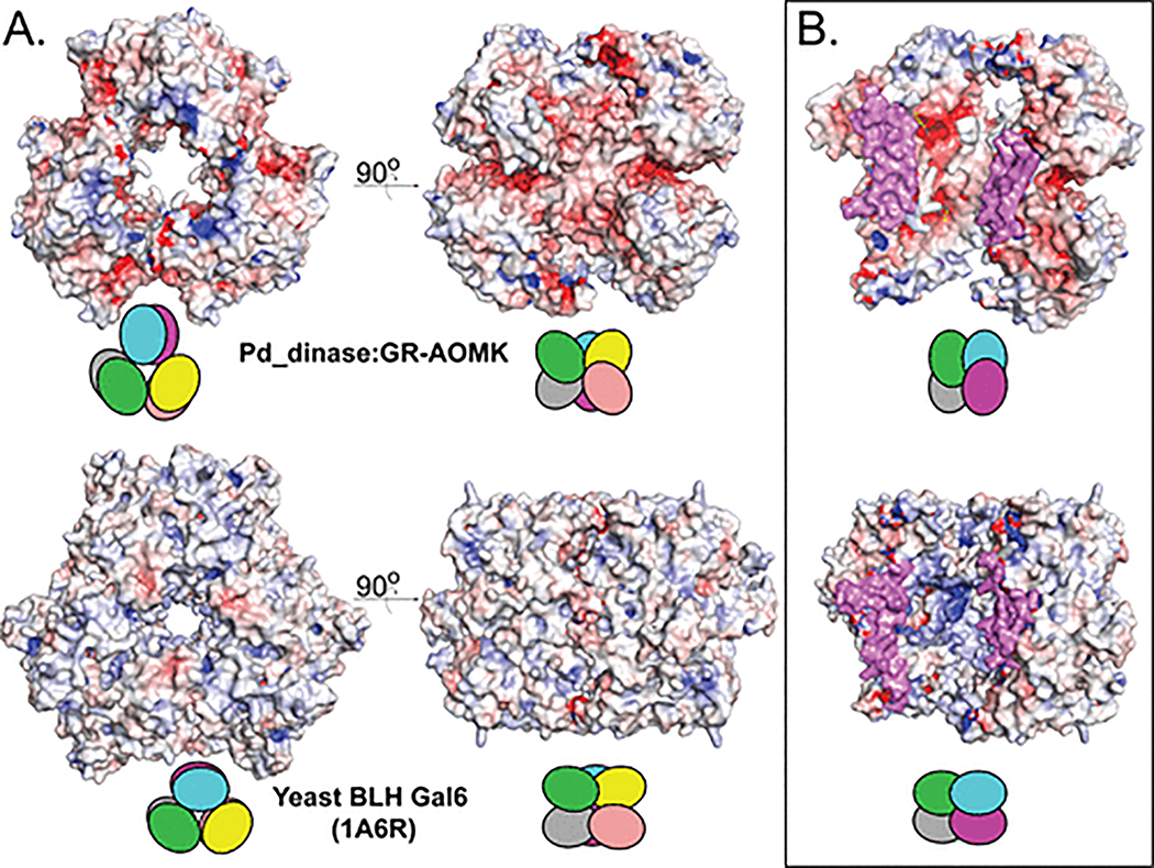
Fig. 5.
Comparison between Pd_dinase and yeast BLH homohexamers. (A) Surface representation and electrostatic potential of Pd_dinase (top) and yeast BLH Gal 6 (bottom) homohexamers accompanied by a schematic to illustrate the position of each chain. (B) Two chains are removed from the homohexamers to exhibit the electrostatic potential of the central channel. The interior of yeast BLH (bottom) is strongly positive while Pd_dinase (top) is slightly negative. The oligomer interface is colored magenta. The electrostatic surface potentials are calculated and colored according to Fig. 3.

