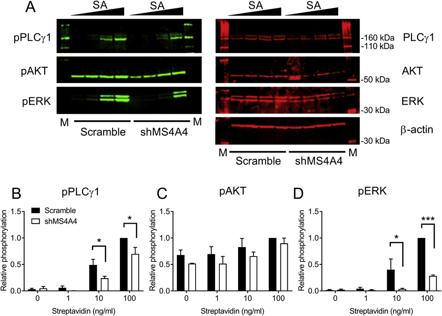
Figure 2. MS4A4A promotes FcεRI-dependent PLCγ1 signaling.

(A) Immunoblots from LAD2 cells transduced with either scramble or shMS4A4A lentiviruses. (B-D) Quantification of PLCγ1 (B), AKT (C), and ERK (D) phosphorylation for scramble or shMS4A4A-transduced LAD2 cells calculated as relative phosphorylation after correction against total protein and normalized to maximal phosphorylation for each phosphoprotein. Data are the mean ± SEM from three independent experiments. *p<0.05, ***p<0.001, ANOVA with Sidak’s posttest.
