Figure 1.

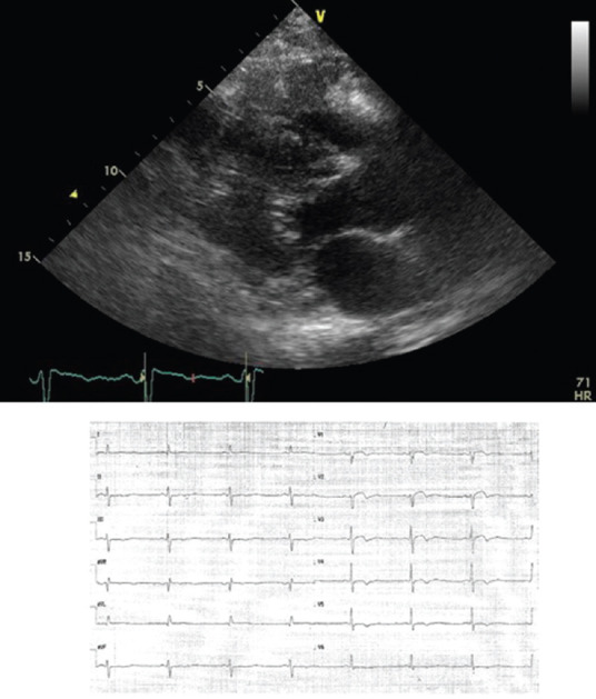
Metastatic angiosarcoma infiltrating the right ventricular wall. Top: 2D-echo long axis view. Down: pseudoischemic anomalies on corresponding leads

Metastatic angiosarcoma infiltrating the right ventricular wall. Top: 2D-echo long axis view. Down: pseudoischemic anomalies on corresponding leads