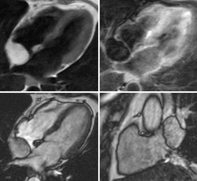
Figure 8.

Lypoma, 60-year old woman, with poor acustic window echocardiogram. Up, cMRI showing rounded capsule formation in right atrium on interatrial septum. Down, hyper-intense in T1 sequence and hypo-intense in fat sat sequences compatible with lypoma (courtesy of Dr. Matteo Gravina)
