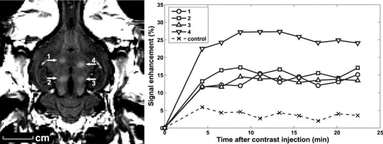
FIGURE 2.
Focal BBB disruption in the rabbit brain using FUS. Left panel, Localized extravasation of Magnevist (MRI contrast agent) in areas of blood-barrier disruption as seen on contrast enhanced T1-weighted brain MRI. Right panel, Change in contrast enhancement over time in the 4 sonicated brain regions and in nonsonicated (control) brain region. Reprinted with permission from Jolesz FA, McDannold N. Current status and future potential of MRI‐guided focused ultrasound surgery. Journal of Magnetic Resonance Imaging. 2008;27(2):9. (c) 2008 Wiley-Liss, Inc.

