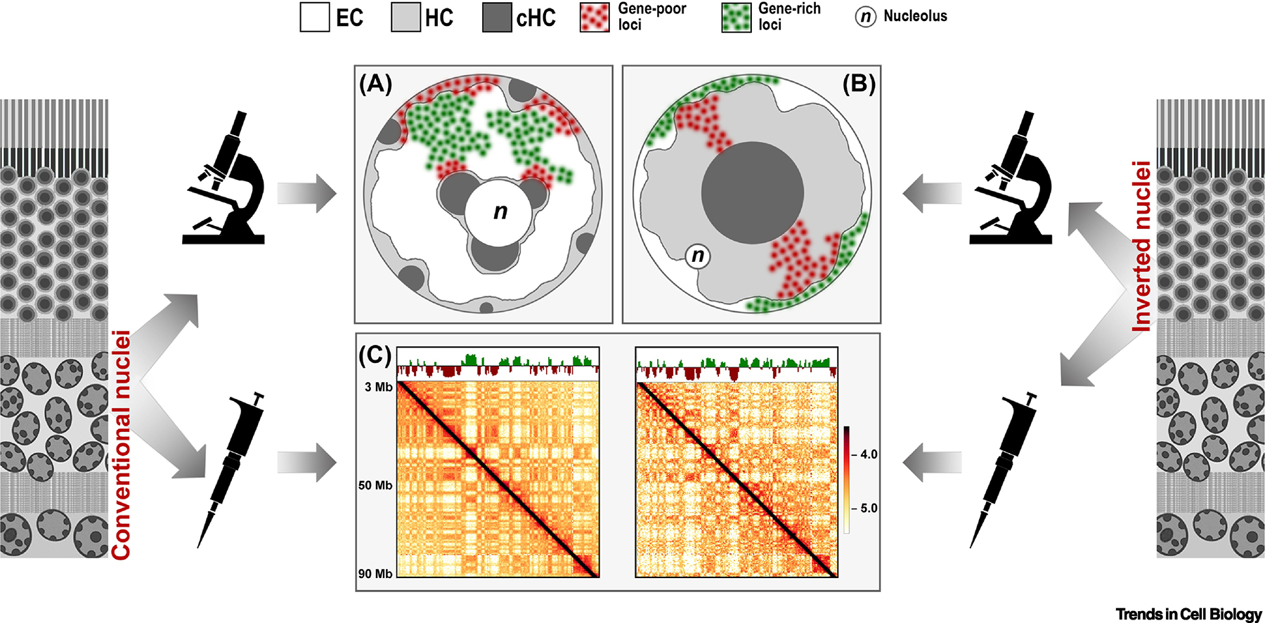
Figure 1. Comparison of conventional and inverted nuclear organization as seen by microscopy (A,B) and Hi-C (C).

Microscopy reveals a striking difference in the nuclear organization of conventional nuclei, e.g., nuclei of non-photoreceptor retinal neurons (A), and inverted nuclei of rod photoreceptors (B) from mouse retina. In conventional nuclei, both types of heterochromatin, HC and cHC, underlie the nuclear envelope and surround the nucleolus (n), whereas EC occupies the nuclear interior. In inverted nuclei, HC and EC exchange their locations: HC is densely packed in the nuclear interior and EC is relocated to the very nuclear periphery. Correspondingly, if genic EC loci (green), easily establish contacts in the nuclear interior of conventional nuclei, contacts between EC loci in inverted nuclei are hampered by the practically two-dimensional organization of the thin layer of peripheral chromatin. In difference to microscopy, bulk Hi-C analysis does not sense the dramatic difference between the two spatial chromatin organizations and in both cases reveals qualitatively similar features, such as chromosome territories, compartments and TADs (C). Shown are Hi-C maps of chromosome 1 in conventional (left) and inverted (right) nuclei.
