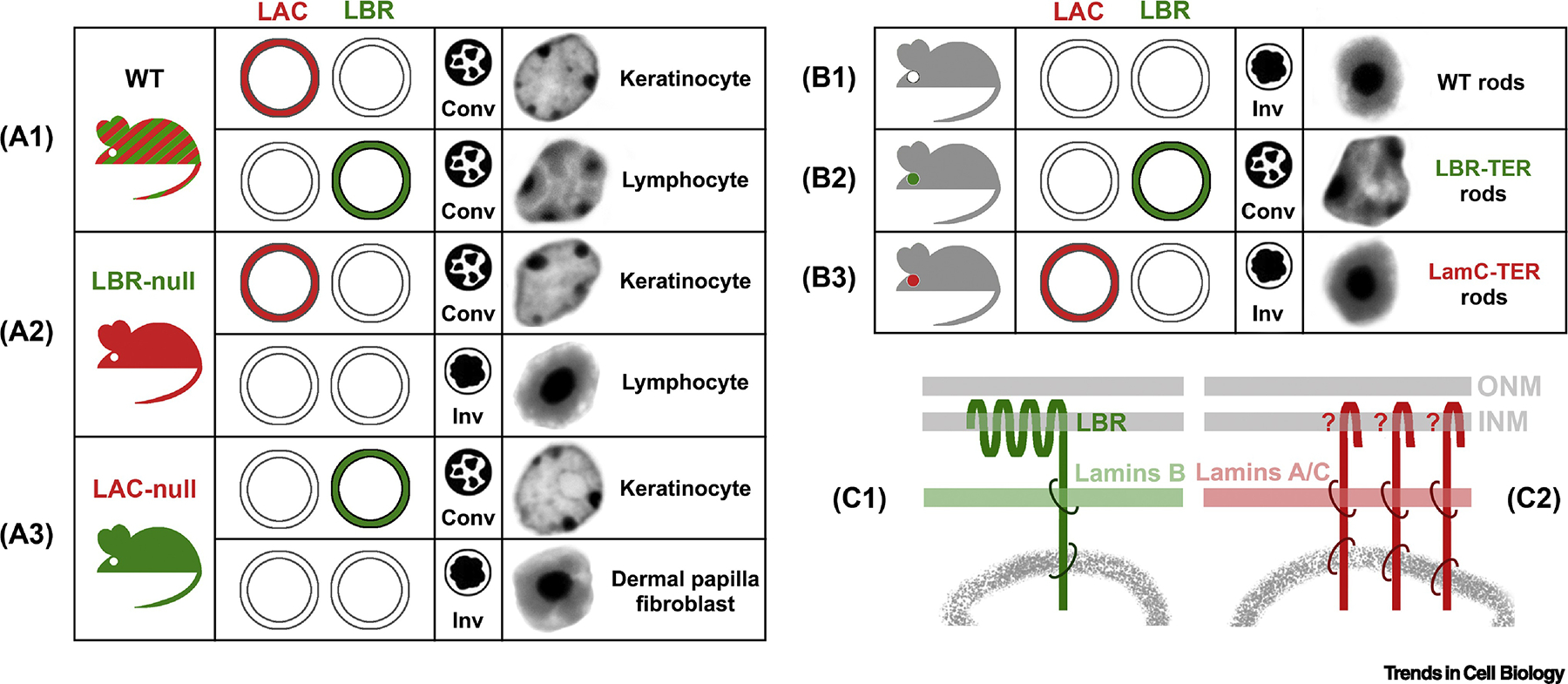
Figure 3. Two major tethers of peripheral heterochromatin.

(A1) LBR and lamin A/C (LAC) are ubiquitously expressed proteins of the nuclear lamina. WT cells either express both proteins or at least one of them and thus have conventional nuclei. (A2) In LBR-null mice, nuclei of those cells that do not normally express LAC, become inverted, e.g., lymphocytes. (A3) In LAC-null mice, nuclei of cells that do not express LBR in WT, become either inverted (e.g, fibroblasts of the dermal papilla) or remain conventional as a result of abnormally upregulated LBR expression (e.g., keratinocytes). (B1) Rods are the only cell type naturally lacking both proteins. (B2) Transgenic expression of LBR in mouse rods effectively counteracts nuclear inversion and restores the conventional architecture in these cells. (B3) Transgenic expression of lamin C is not sufficient for restoration of the conventional architecture, suggesting that LAC needs partners for heterochromatin binding, which are not expressed in mouse rods. (C) Schematics of the two major types of peripheral heterochromatin tethers. LBR-dependent tether (C1) binds B-lamins and peripheral heterochromatin. LAC-dependant tether (C2) includes transmembrane proteins (marked with “?”) binding both LAC and heterochromatin. conv, conventional nuclei; inv, inverted nuclei; LBR-TER, transgenic rods expressing ectopic LBR; LamC-TER, transgenic rods expressing ectopic lamin C; nuclei of exemplified cells on (A) and (B) are shown as inverted greyscale images of DAPI staining; ONM, outer nuclear membrane; INM, inner nuclear membrane.
