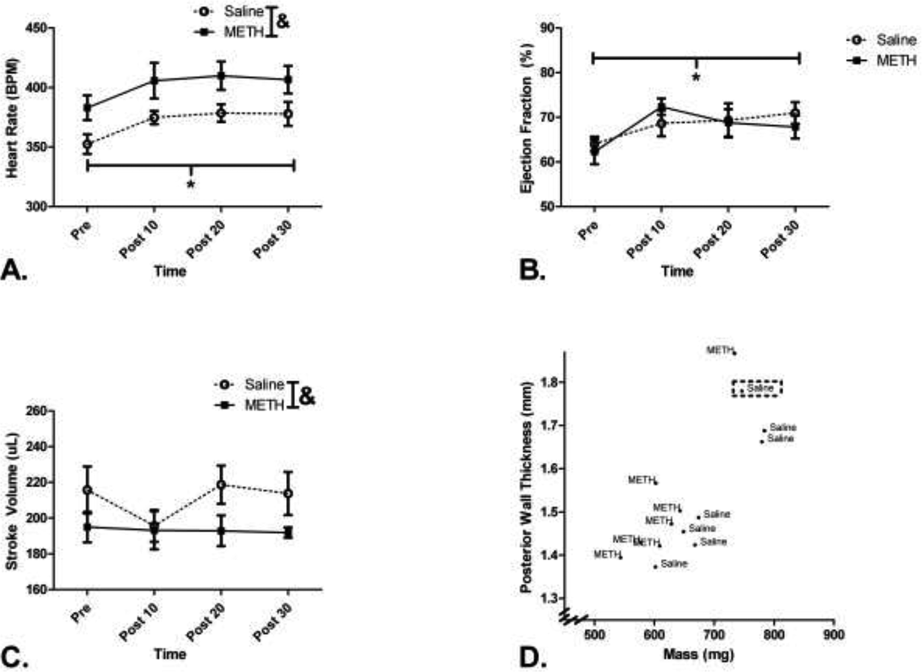
Figure 2.

Echocardiogram measures as assessed on the day following the 9th self-administration. Basal cardiac parameters were assessed followed by a 1 mg/kg injection of METH. Cardiac parameters were then reassessed every 10 min after the injection. A. METH self-administering animals had elevated heart rates compared to saline animals, but both groups’ heart rate increased following the METH challenge. B. Ejection fraction increased following the METH challenge in both groups. C. METH self-administering animals had reduced stroke volumes compared to saline self-administering animals. D. A scatter plot of average left ventricular volume versus average diastolic left ventricular posterior wall thickness following 9 d of self-administration. Linear discriminate analysis was performed using these parameters and cross-validated to predict group assignment. The animal that was misclassified during the training of the linear discriminate analysis is outlined with dashed markers. & p<0.05 METH vs. Saline. *p<0.05 Pre vs. Post 10, Post 20, Post 30.
