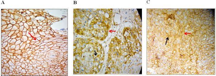
Figure 2.
Expression of Beta-Catenin in Different Oral Mucosal regions; (A), Beta - catenin in PEH showing intense membranous staining (red arrow) throughout the epithelium; (B), Beta - catenin in WDOSSC showing membranous (black arrow) and cytoplasmic (red arrow) staining; (C)Beta-catenin in PDOSCC showing dispersed altered cytoplasmic (black arrow) and nuclear staining (red arrow) throughout the cancerous islands (immunohistochemistry staining, original magnification 400x).

