Figure 4.
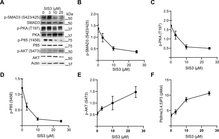
SMAD3 inhibition restores PI3K activity. A, murine CD4+ T cells were pretreated with various doses of SIS3 inhibitor for 2 h and activated through the TCR, CD28, and with 10 ng/ml TGF-β for 10 min. Immunoblotting was performed for various phosphoproteins. One representative blot from two independent experiments is shown. B–E, immunoblots from panel A were quantitated by densitometry and normalized to total protein abundance for (B) p-SMAD3 (Ser-423/425), (C) p-PKA (Thr-197), (D) p-P85 (Ser-458), and (E) p-AKT (Ser-473). Shown are mean ± S.D. from two independent experiments per data point. F, lipids were extracted from activated T cells and mass ELISA were utilized to measure the levels of PtdIns(3,4,5)P3 as a function of SIS3 dose (n = 2 per data point).

