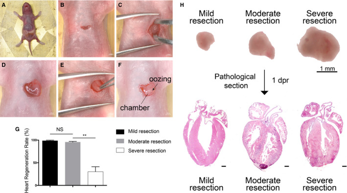
FIGURE 1.

Thoracotomy optimization in neonatal mouse apical resection model. A, Mouse was fixed on the operating table in the supine position. B, The skin was incised for 1 cm long. C, The heart was exteriorized out of the chest cavity with the help of a forceps. D, The heart was immobilized by surrounding chest tissues. E, Clamping the heart out of the chest damaged the heart tissue mechanically. F, The ventricular chamber was exposed with one cut. Exposure of the chamber was the landmark of successful resection. G, Heart regeneration rate of different sizes of resection. n = 20 each. Data are mean ± SEM. NS, no statistical significance, **P < .01. H, Illustration of different cut sizes. Microscopic images (upper) depict HE staining (down). Mild resection (<0.5 mm) was too small to properly expose the chamber. Severe resection (>1.5 mm) could easily expose the chamber but with a lower regeneration rate. Moderate resection (about 1 mm) was the perfect size to replicate the model. 1dpr, 1 day post‐resection
