Figure 3.

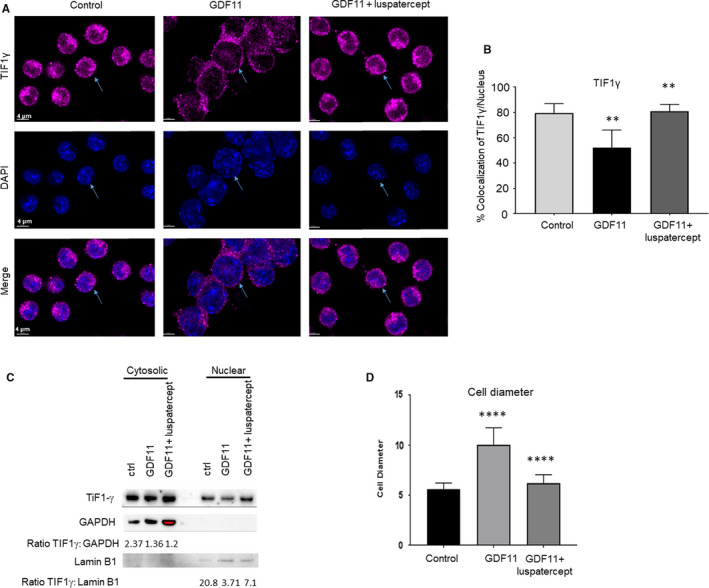
Smad2/3‐pathway overactivation reduces, and luspatercept co‐treatment promotes, nuclear localization of TIF1γ in MEL cells. A, Immunofluorescence microscopy showing effect of GDF11 (100 ng/mL, 24 h) alone or in combination with luspatercept (1 µg/mL) on TIF1γ levels in DMSO‐pretreated MEL cells (control). Representative images depict TIF1γ (magenta/AF647). TIF1γ levels were lower overall and localized to a lesser degree in the nucleus under GDF11‐treated conditions compared with control and luspatercept co‐treatment. Scale bar, 4 µm. B, Percentage of cells with nuclear TIF1γ localization. Data are means ± SEM (n = 3 images per group), *P < .05 vs. DMSO or co‐treatment. C, Western blot analysis showing effect of GDF11 (100 ng/mL) alone or in combination with luspatercept (1 μg/mL) on TIF1γ protein expression in cytosolic and nuclear fractions of MEL cells pretreated with 2% DMSO (control) to induce differentiation. GAPDH served as loading control for cytosolic extracts, and LaminB1 is used as nuclear protein loading control for nuclear extracts. D, Cell diameter measurement from panel A. Data are means ± SEM (n = 10 randomly selected cells per group), ****P < .0001 vs. control or co‐treatment. Note larger size of GDF11‐treated cells compared to control (indicated by arrowheads)
