
Figure 8.
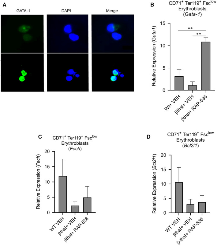
GATA‐1 levels are restored in β‐thalassaemic bone marrow cells by luspatercept in vitro and in erythroblasts of Hbbth1/th1 β‐thalassaemic mice by RAP‐536 in vivo. (A) Immunofluorescence microscopy showing effect of luspatercept (1 µg/mL, 48 h) on nuclear levels of GATA‐1 in cultured bone marrow cells from Hbbth1/th1 β‐thalassaemic mice. Images depict GATA‐1 (green/AF488) and DAPI‐labelled nuclei (blue). Expression of Gata1 detected by qPCR in (B) sorted CD71highTER119+FSClow splenic erythroblasts. Expression of (C) Fech, (D) Bcl2l1 by qPCR in sorted splenic erythroblasts from wild‐type mice and β‐thalassaemic mice treated with a single dose of RAP‐536 (30 mg/kg) or vehicle for 16 h. Data are means ± SEM (n = 3 mice per group). ** P < .01 vs β‐thal + RAP‐536 by one‐way ANOVA with post hoc Tukey HSD test. Data B‐F are normalized against GAPDH