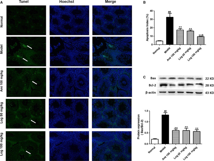
FIGURE 4.

Loganin relieved apoptosis in the testes of KK‐Ay mice detected by TUNEL staining. A, TUNEL‐positive cells (white arrows) in representative images are shown in green (200 × magnification). Cell nucleus was subjected to Hoechst staining. B, Quantification of apoptotic cell percentage using Image J software. C, Western blot analyses of Bax and Bcl‐2 protein expressions in testis homogenate. Bax/Bcl‐2 ratio was calculated and analyzed. Bars represent mean ± SD, n = 3. Significance: ## P < 0.01 vs normal group; *P < 0.05, **P < 0.01 vs model group; & P < 0.05 vs aminoguanidine group
