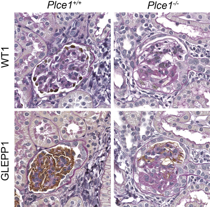
Fig. 5.

Representative light microscopy images of glomeruli from phospholipase C-ε1 (Plce1)+/+ and Plce1−/−mice treated with DOCA + salt + uninephrectomy + norepinephrine. Focal glomerulosclerosis was noted within Plce1−/− mice treated with this protocol. Note the lack of Wilms’ tumor-1 (WT1) and receptor-type protein-tyrosine phosphatase O (GLEPP1) staining within the Plce1−/− glomerulus, consistent with focal podocyte depletion. Magnification: ×40.
