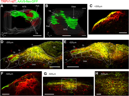
Figure 17.
Brainstem terminations of vagal TRPV1-expressing afferents labeled by unilateral intraganglionic injection of AAV9-flex-GFP into TRPV1-tdT. AAV-mediated GFP expression (green, enhanced by α-GFP immunoreactivity) is compared with ROSA26-mediated tdTomato expression (red, native) in coronal sections of the medulla. A, B, 3D reconstruction of medulla along entire rostral–caudal axis. A, Rostral aspect. B, Dorsal aspect. C–H, Coronal sections of medulla from rostral to caudal, with labeling for the position relative to obex. C, Pa5 at +300 μm (relative to obex). D, E, nTS at −200 μm (D) and −520 μm (E). The following structures are identified: area postrema (AP), DMX, SolC (C), SolCe (Ce), SolDL (DL), SolG (G), SolIM (IM), SolM (M), SolV (V), SolVL (VL), and TS. F, Pa5 at −520 μm. G, H, nTS at −820 μm. H, High-magnification image of SolC area identified by white box in G. Scale bars: A, B, 1 mm; C–G, 200 μm; H, 40 μm.

