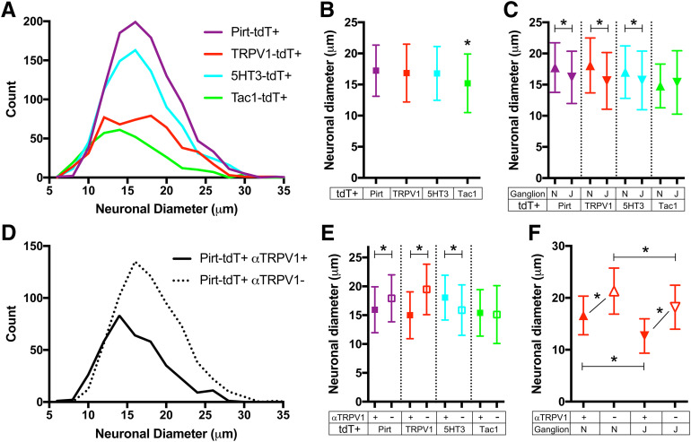
Figure 3.
Neuronal diameters of tdTomato+ vagal neurons from Pirt-tdT, 5-HT3-tdT, TRPV1-tdT, and Tac1-tdT. A, Histogram of neuronal diameter of tdTomato+ vagal neurons from Pirt-tdT (n = 1040 neurons), 5-HT3-tdT (n = 850 neurons), TRPV1-tdT (n = 501 neurons), and Tac1-tdT (n = 330 neurons). B, Mean ± SD neuronal diameter of tdTomato+ vagal neurons from Pirt-tdT, 5-HT3-tdT, TRPV1-tdT, and Tac1-tdT. C, Mean ± SD neuronal diameter of tdTomato+ nodose neurons (upward triangles) compared with jugular neurons (downward triangles) from Pirt-tdT, 5-HT3-tdT, TRPV1-tdT, and Tac1-tdT. D, Histogram of neuronal diameter of tdTomato-expressing neurons with α-TRPV1 immunoreactivity (Pirt-tdT+αTRPV1+, black line) and tdTomato-expressing neurons without α-TRPV1 immunoreactivity (Pirt-tdT+αTRPV1−, dotted line) from Pirt-tdT. E, Mean ± SD neuronal diameter of tdTomato+ vagal neurons with (filled squares) and without (open squares) α-TRPV1 immunoreactivity from Pirt-tdT, 5-HT3-tdT, TRPV1-tdT, and Tac1-tdT. F, Mean ± SD neuronal diameter of tdTomato+ nodose (upward triangles) and jugular (downward triangles) neurons with (filled squares) and without (open squares) α-TRPV1 immunoreactivity from TRPV1-tdT. *Denotes significant difference (p < 0.05, ANOVA with Sidak’s multiple comparisons).

