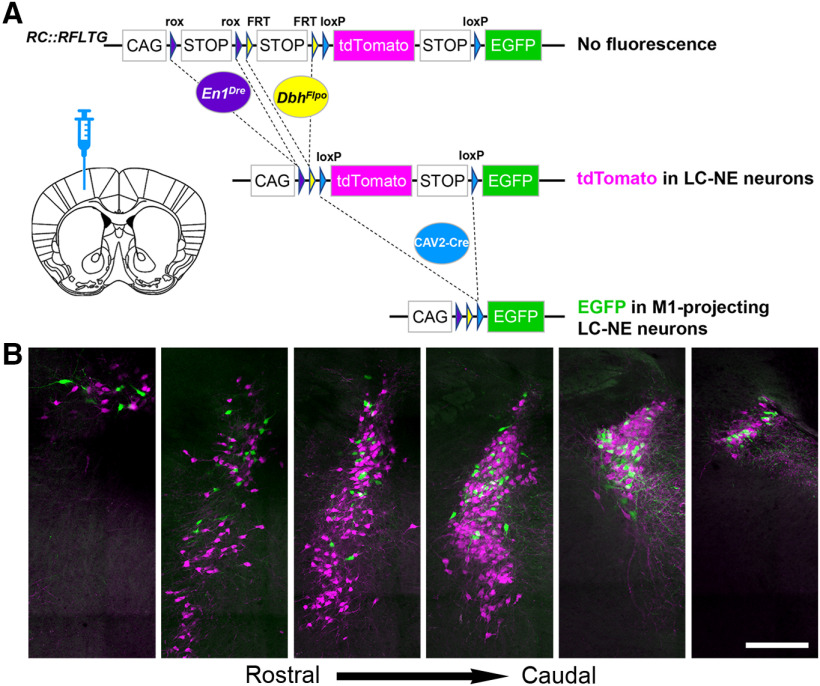
Figure 4.
Location of motor cortex-projecting noradrenergic neurons within the LC. A, Coronal schematic of mouse forebrain section showing position of CAV2-Cre injection. B, Representative coronal sections through the rostrocaudal extent of the ipsilateral LC from a TrAC-LC mouse (40-μm virtual sections from PACT-cleared tissue) showing distribution of EGFP-labeled (green) and tdTomato-labeled (magenta) neurons. Scale bar, 200 μm.

