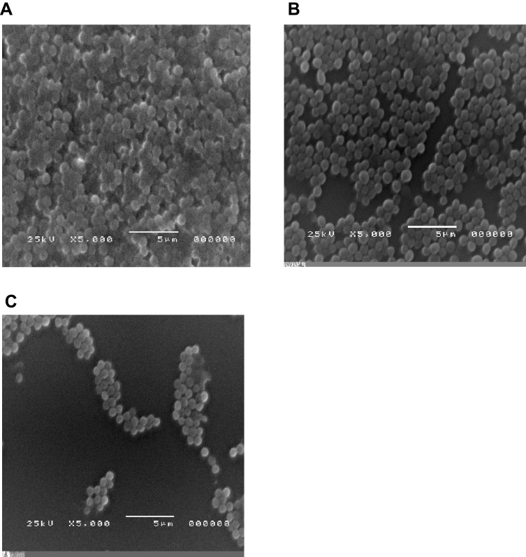
Figure 3.
Visualization of S. aureus biofilms formed in the presence of subinhibitory concentration of free ciprofloxacin and ciprofloxacin-loaded niosomes, using an electron microscope. (A) Untreated culture (positive control), (B) culture in the presence of 1/8 MIC of ciprofloxacin, (C) culture in the presence of 1/8 MIC of ciprofloxacin-loaded niosomes.

