
Figure 2.
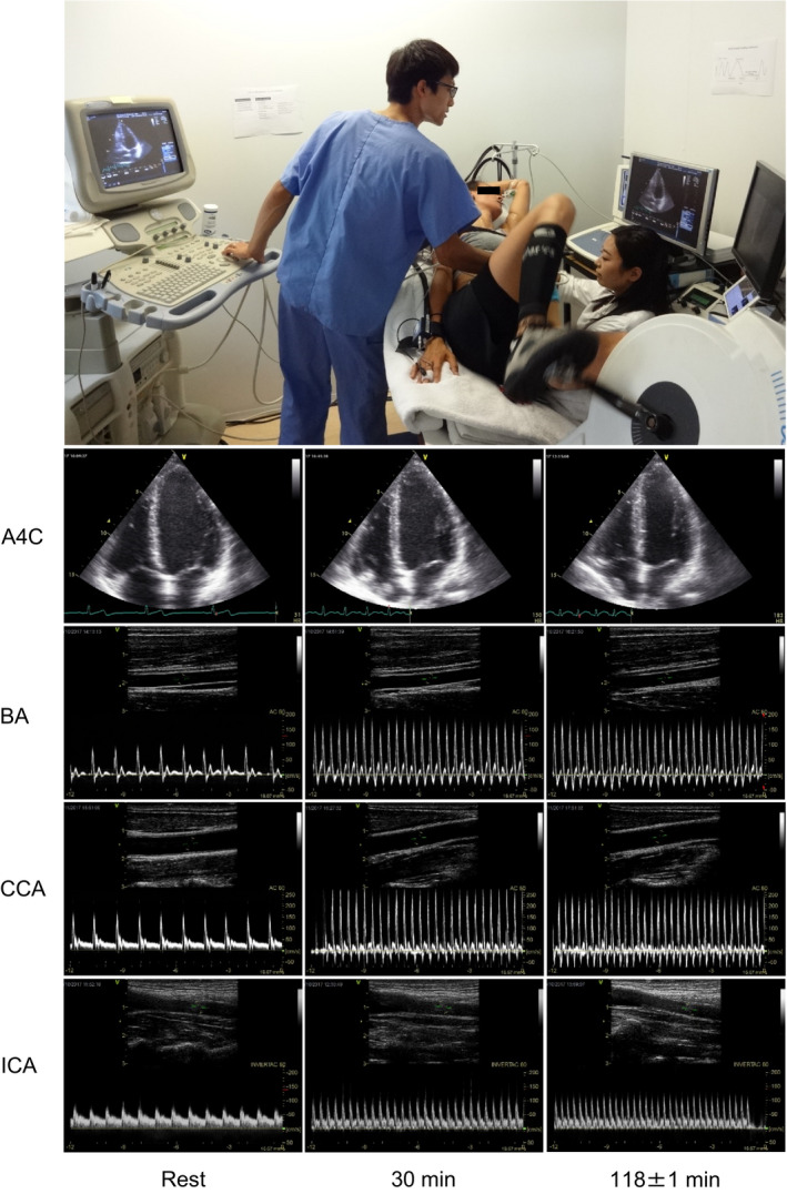
Examples of echocardiographic and Doppler ultrasonographic assessments during prolonged submaximal semi‐recumbent cycling. The photo depicts one of the participants in the study. Representative images of apical 4‐chamber view at end diastole (A4C) and vessel and blood velocity recordings at the brachial artery (BA), common carotid artery (CCA), and internal carotid artery (ICA), at rest and during exercise (30 and 118 ± 1 min) in the progressive dehydration trial are shown