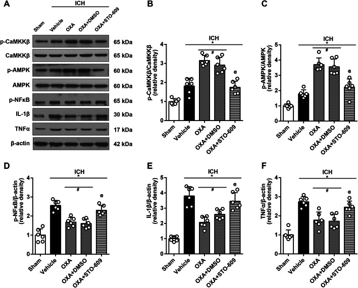
Fig. 7.
Western blot analysis showing anti-inflammatory effects of OXA. a Representative protein bands and the quantitative analysis of b ratio of p-CaMKKβ/CaMKKβ and c ratio of p-AMPK/AMPK at 24 h after ICH. The expression of p-CaMKKβ and p-AMPK was upregulated after ICH and was further elevated by OXA. This effect was reversed with STO-609 (p-CaMKKβ inhibitor). Consequently, the inflammatory factors including d p-NFκB, e IL-1β, and f TNFα were increased after ICH and were significantly suppressed by OXA. Consistently, this effect was reversed with STO-609. One-way ANOVA, *p < 0.05 vs. sham group, #p < 0.05 vs. vehicle group, @p < 0.05 vs. OXA group. Error bars represent mean ± SD, n = 6 per group

