
Figure 3.
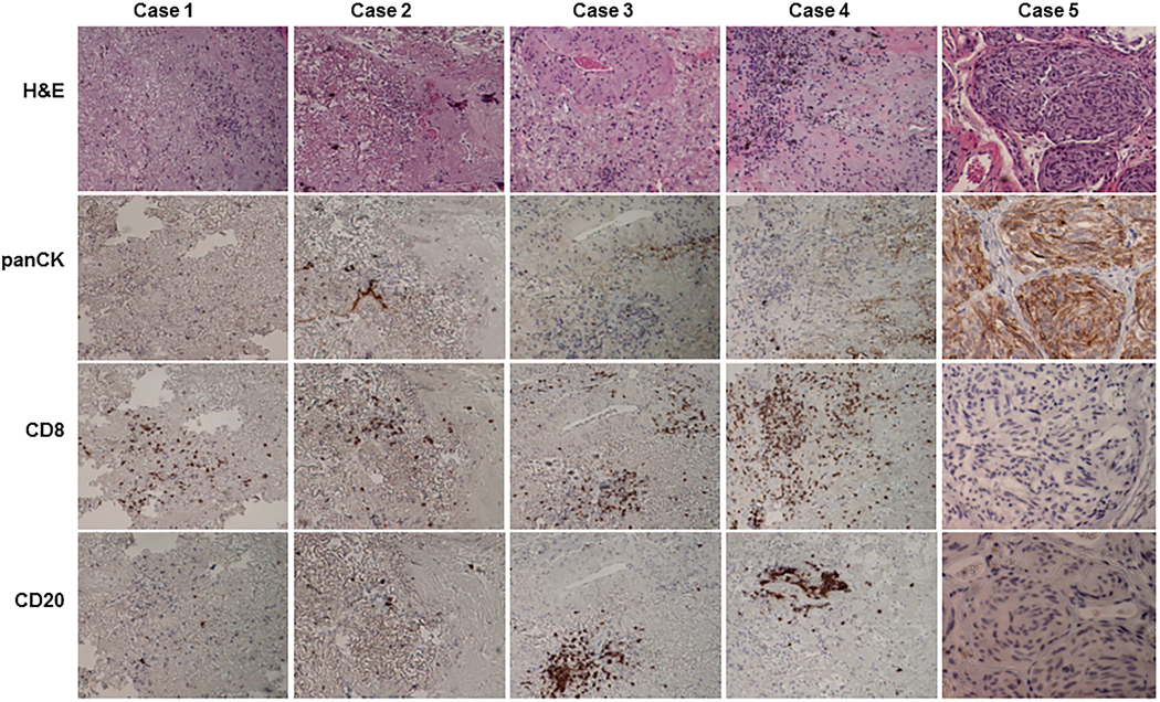
Histological characterization of residual tumor tissue that did not form PDXs. Residual tumor tissues were harvested at 12 months after implantation from mice that showed no signs of PDX growth. On hematoxylin and eosin staining, the residual tumors from cases 1 to 4 were composed mainly of fibrotic tissues with scattered or clustered inflammatory cells. Case 5 had viable tumor cells. Immunohistochemical staining showed that inflammatory cells were human CD8+ or CD20+ cells, whereas cancer cells were positively stained for pan-cytokeratin (panCK).