Figure 2.

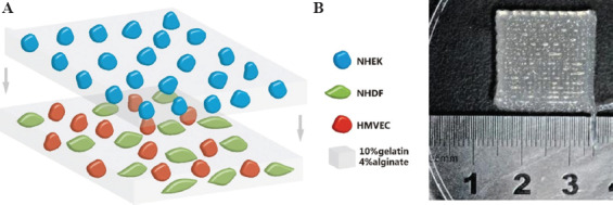
(A) Schematic diagram of transplantable printed skin. The top layer consisted of keratinocytes and gel and the bottom layer fibroblasts, microvascular endothelial cells and gel. (B) A macroscopic image of the printed skin graft.

(A) Schematic diagram of transplantable printed skin. The top layer consisted of keratinocytes and gel and the bottom layer fibroblasts, microvascular endothelial cells and gel. (B) A macroscopic image of the printed skin graft.