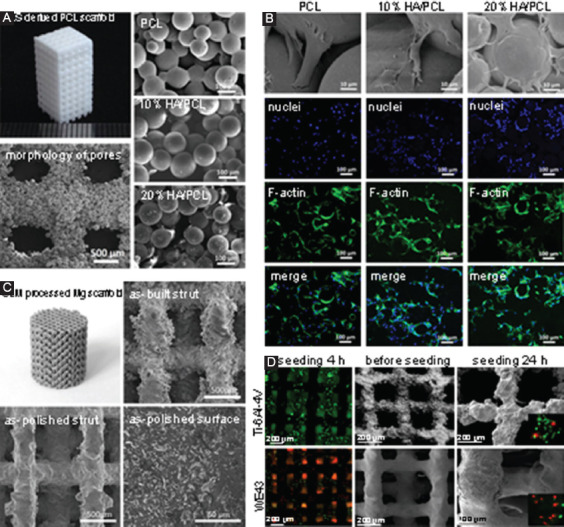
Figure 6.

(A) Selective laser sintering (SLS)-derived poly(ε-caprolactone) scaffold, with scanning electron microscope (SEM) images showing the morphology of pores and well-connected microspheres[99]. (B) In vitro evaluation of SLS-derived scaffolds, with SEM and confocal images showing the morphologies of adherent mesenchymal stem cells on the scaffolds after culturing for 12 h. (C) Selective laser melting (SLM) processed Mg-based scaffold (WE43), with SEM images showing the surface morphology and microstructure[124]. (D) In vitro evaluation of SLM-derived WE43 and Ti-6Al-4V scaffolds. Fluorescent optical images showed the morphologies of MG 63 cells on scaffolds, in which live cells were stained in green, whereas dead cells were stained in red.
