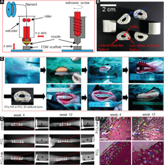
Figure 7.

(A) A diagram for fused deposition modeling (FDM) process. (B) The FDM-derived poly(ε-caprolactone)/hydroxyapatite (PCL/HA) scaffolds[139]. (C) The implantation of FDM-derived PCL/HA scaffolds. (D) X-ray images of the goat legs after implantation for 4 and 12 weeks, in which the arrows mark the bone defect edges. (E) Histological imagines showing the interfaces between the scaffolds and the surrounding tissue. AB represents artificial bone, FB represents natural goat femur bone, and NB represents new bone. The scaffolds were filled with new bone after 12 weeks’ implantation.
