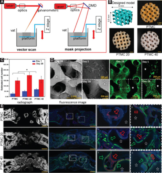
Figure 9.

(A) A diagram for two irradiation types for stereolithography (SLA), including vector scan and mask projection. (B) Poly(trimethylene carbonate) (PTMC) scaffolds fabricated by SLA with various hydroxyapatite (HA) contents, with PTMC20 and PTMC40 containing 20 and 40 wt.% HA, respectively[160]. (C) In vitro cell culture on scaffolds, with (D) scanning electron microscope and fluorescence images showing the different cell morphologies on the scaffold. (E) Contact radiographs of the defects combined with fluorescence images showing the newly formed bone after implantation for 2 weeks (in green) and 4 weeks (in red).
