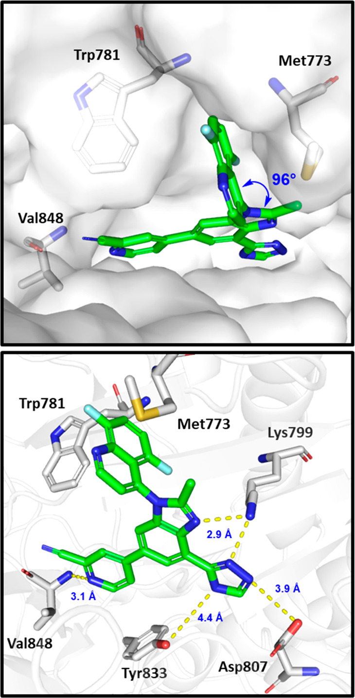
Figure 2.

Docked pose of (P)-14 in the homology model of PI3Kβ built from the cocrystal structure of (P)-19 bound to PI3Kδ (pdb: 6DGT).19 Some residues have been removed for clarity, and yellow dashed lines show hydrogen bond contacts between the inhibitor and the protein.
