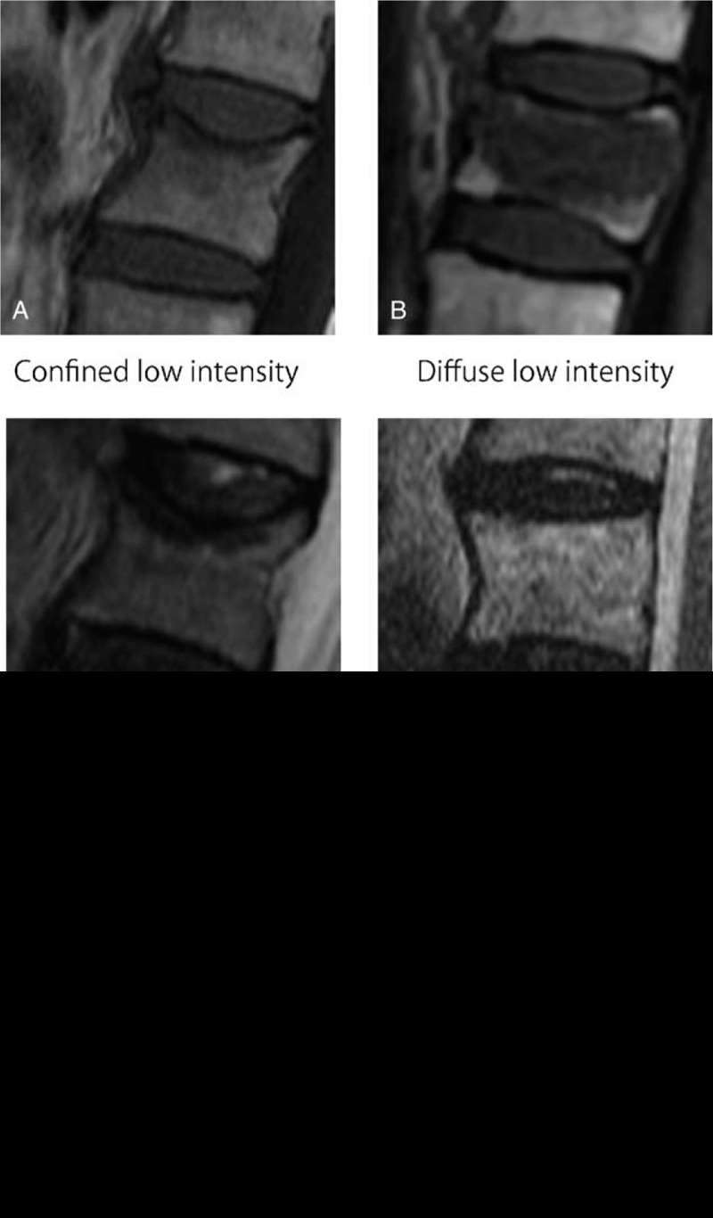
Figure 1.

Classification of OVFs on T1-weighted images. A, Confined low-intensity image. B, Diffuse low-intensity image. Classification of OVFs on T2-weighted images. C, Confined low-intensity image. D, Diffuse high-intensity image. E, Diffuse low-intensity image. F, Fluid-intensity image. OVF indicates osteoporotic vertebral fractures.
