Figure 5.
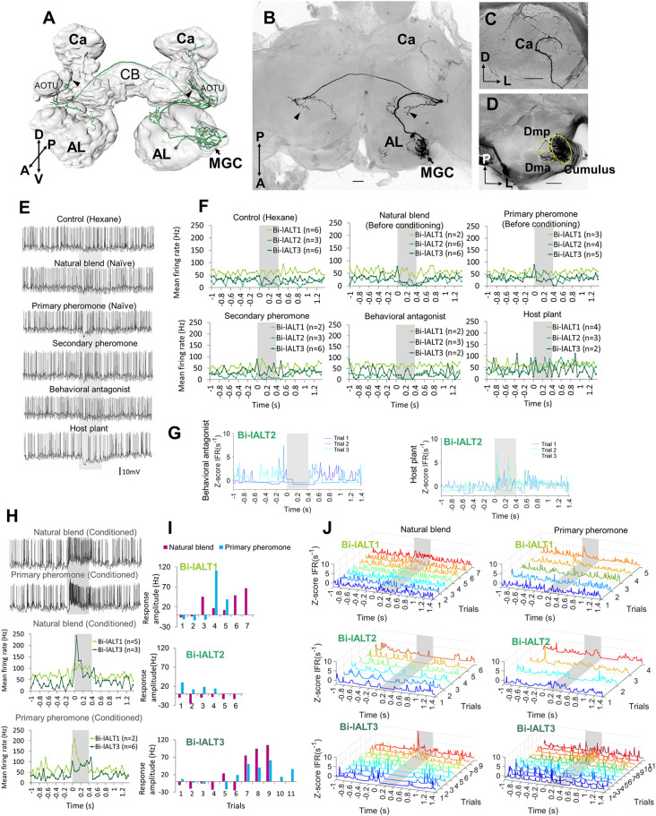
Morphology and electrophysiology of bilateral lateral-tract neurons with restricted projections in the protocerebrum (Sub-type I). (A) 3D reconstruction of one neuron (Bi-lALT3) in dorsal-frontal view, demonstrating projections to ipsilateral and contralateral column (arrowheads), and ipsilateral calyx (Ca). (B–D) Confocal images of Bi-lALT3. The fiber projecting to the ipsilateral Ca is shown in (C). Arborizations in the antennal lobe (AL) are shown in (D), including dendrites restricted to the MGC, in which the cumulus is considerably stronger innervated than dma and dmp. (E) Spiking activity of one neuron (Bi-lALT3) during the first-trial application of each stimulus (i.e., naïve). (F) Mean spiking frequencies during repeated trials in the three Sub-type I bilateral neurons before induced response appeared, i.e., first three trials for Bi-lALT1 and first five trials for Bi-lALT3 (bin-size: 50 ms, n: number of repeated trials). (G) Traces of instantaneous firing rates of one neuron (Bi-lALT2) showing an inhibitory response to the behavioral antagonist and an excitatory response to the host plant. (H) Induced response in neuron Bi-lALT3. Upper part: spiking activities of the neuron during the 7th application of natural blend and primary pheromone, respectively. Lower part: spiking frequencies to the same stimuli during 4th–7th trials in neuron Bi-lALT1 and 7th–11th trial in neurons Bi-lALT3 (bin-size: 50 ms, n: number of repeated trials). (I) Induced responses are present in two of the three sub-type I bilateral neurons, i.e., Bi-lALT1 and Bi-lALT3 (no effect in Bi-lALT2). (J) Traces of instantaneous firing rates illustrating variable responses to the natural blend and the primary pheromone across repeated trials in the same two Sub-type I bilateral lALT neurons, Bi-lALT1 and Bi-lALT3. AOTU, anterior optic tubercle; CB, central body. A, anterior; L, lateral; M, medial; P, posterior; V, ventral. AL, antennal lobe; Ca, Calyces of the mushroom body; CB, central body. A, anterior; L, lateral; M, medial; P, posterior; V, ventral. Scale bars, 50 μm. Gray bar, the duration of the stimulus (400 ms).

