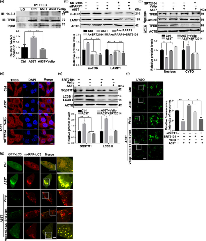
Figure 3.

Veliparib rescues the autophagy flux via activating SIRT1 in α‐synucleinA53T model of Parkinson's disease. (a) SN4741 cells were transfected with Adenovirus‐Ctrl/A53T and further treated with Veliparib (10 μM, 12 hr). The interaction between 14‐3‐3 and TFEB was tested in TFEB Immunoprecipitates. Means ± SEM, n = 3. (b) Representative immunoblots and quantification of the levels of mTOR, LAMP1 in SN4741 cells with different treatments. Mean ± SEM, n = 3. (c) Representative immunoblots and quantification of the levels of TFEB from nuclear and cytoplasmic fractions in SN4741 cells with different treatments. (d) Immunostaining of TFEB (red) and DAPI (blue). Scale bars, 7 μm. (e) Representative immunoblots and quantification of the levels of SQSTM1, LC3B in SN4741 cells with different treatments. Mean ± SEM, n = 3. (f) Representative images and quantification of Lysosomes (green) in SN4741 cells transfected with Adenovirus‐Ctrl/A53T and further treated with Veliparib (10 μM, 12 hr), SRT2104 (10 μM, 12 hr), Veliparib plus EX527 (10 μM, 12 hr). Scale bars, 10 μm. (g) Representative images of LC3 puncta in SN4741 cells transfected with GFP‐mRFP‐LC3B plasmid. Scale bars, 5 μm (the statistical significantly was analyzed by unpaired Student's t test or one‐way ANOVA, *p < .05, **p < .01, and ***p < .001). TFEB, transcription factor EB
