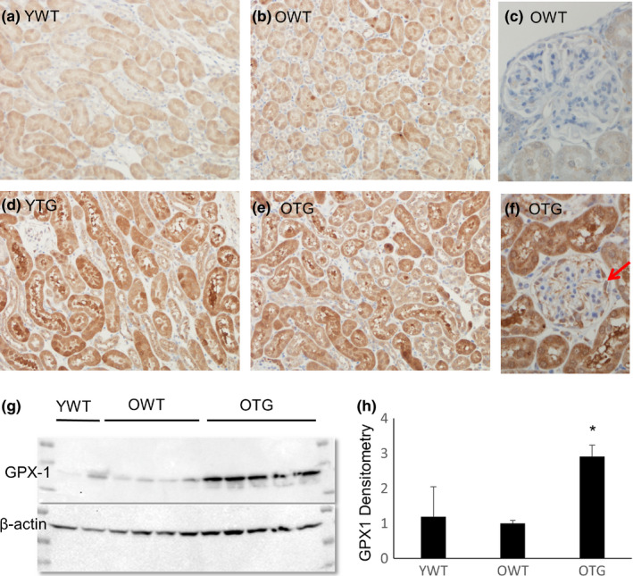
FIGURE 1.

Levels of GPX1 protein in kidneys of wild‐type (WT) and GPX1 transgenic (TG) mice, at young (Y, 4–5 months old) and old (O, 21–23 months old) ages. (a–f) Immunohistochemistry for GPX1. The GPX1 TG was overexpressed in tubular epithelial cells (d, e) and podocytes (f, arrow). (g, h) Immunoblots showing an ~3‐fold increase of GPX1 in kidneys from old TG mice compared with old WT mice. N = 4–5. *p < .05
