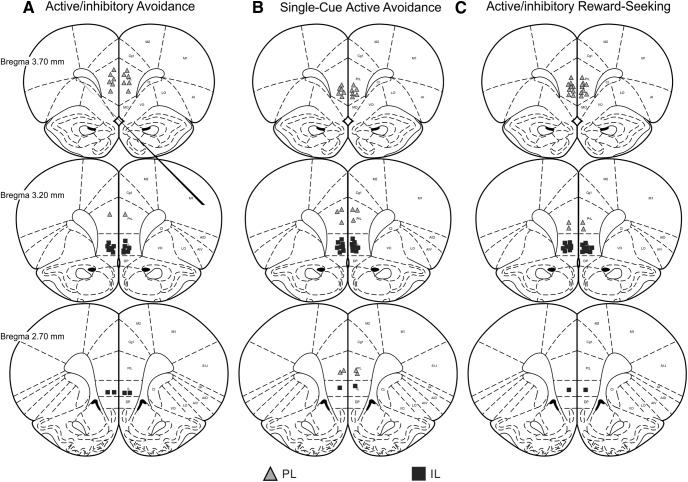
Figure 2.
Histology. Schematic of coronal sections displaying the ventral extent of acceptable microinfusions in the PL (gray triangles) and IL (black squares). Placements are shown for rats in (A) active/inhibitory avoidance, (B) active avoidance, and (C) active/inhibitory reward-seeking experiments.

