. 2020 Jun 11;2(3):e200213. doi: 10.1148/ryct.2020200213

Figure 2:

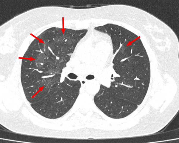
Example of indeterminate CT imaging features for COVID-19 in a 36-year-old female patient. Chest CT image shows bilateral multifocal ground-glass opacities (arrows), which were mainly located in the right upper lobe. There was no posterior part/lower lobe predilection, and there was also no peripheral/subpleural distribution of lung abnormalities.

Example of indeterminate CT imaging features for COVID-19 in a 36-year-old female patient. Chest CT image shows bilateral multifocal ground-glass opacities (arrows), which were mainly located in the right upper lobe. There was no posterior part/lower lobe predilection, and there was also no peripheral/subpleural distribution of lung abnormalities.