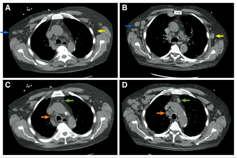
Figure 1. CT scan of the chest.
Increased bilateral axillary adenopathies (blue and yellow arrows) are smaller in the previous CT scan done 10 months prior (A) when compared to the new scan (B). Similarly, increased mediastinal adenopathies are evident (green and orange arrows) when comparing an old scan (C) with the newer study (D)
CT: computed tomography

