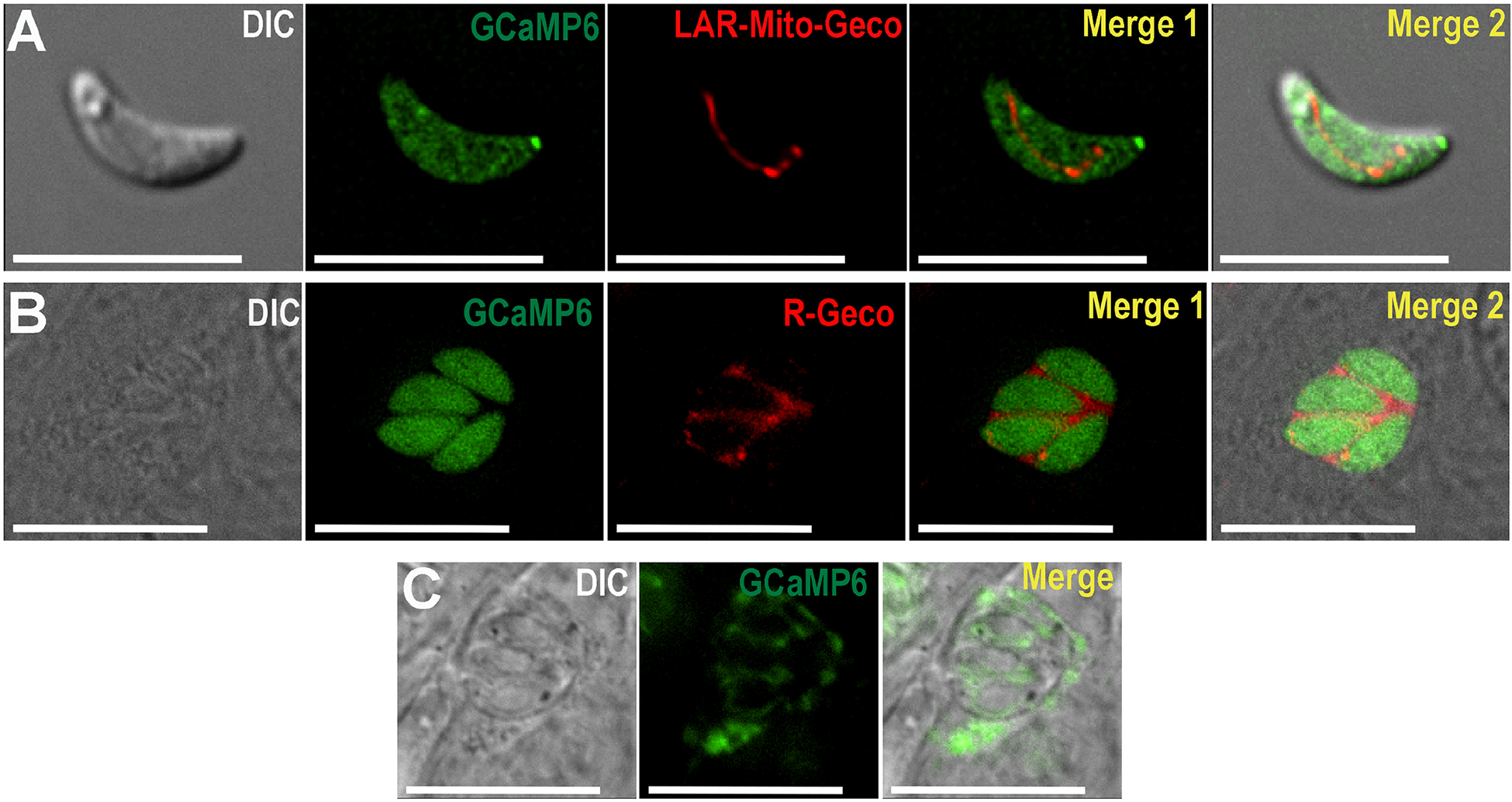
Figure 1: Images of Toxoplasma gondii parasites expressing GCaMP6 in the cytoplasm, mitochondria and parasitophorous vacuole.

A, Live cell imaging of extracellular tachyzoites of the RH strain expressing GCaMP6f (pTUBGCaMP6f) (green) in the cytosol and LAR-Geco (pDT7S4H3-SOD2-Mito-LAR-Geco) in the mitochondria (red). B, Live cell imaging of intracellular parasites expressing GCaMP6f (pTUBGCaMP6f) in the cytosol (green) and R-Geco (ptub_SAG1-IEα-R-GECO_dhfr_sag1CATsag1) in the PV (red). C, Live cell imaging of intracellular RH parasites expressing GCaMP6f (pDT7S4H3-P30-GCaMP6f) in PV.
