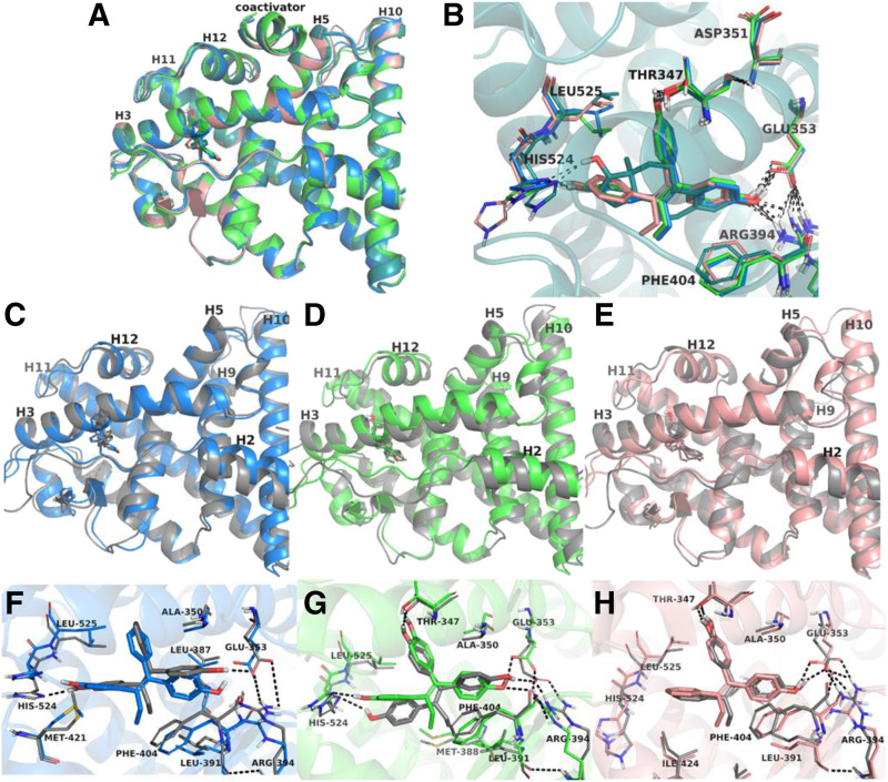
Fig. 9.
Molecular dynamics simulations of the wild-type ER LBD with TPE derivatives. Experimental structures of ER-LBD co-crystallized with E2 (teal), Z2OHTPE (blue), 3OHTPE (green), and BPTPE (pink) are superimposed (A), and the contacts between the ligands and critical amino acids of the binding site are shown (B). For each ligand-receptor complex snapshots taken from the MD trajectory (colored in gray) are overlaid with their experimental structures (C–E, same color code as in A). Close views of the ER binding pocket with Z2OHTPE (F), 3OHTPE (G), and BPTPE (H) show small variations between the experimental structures and the representative conformations extracted from the MD simulations. The same color code is used in pictures (C–H); MD snapshots are colored in gray, whereas the experimental structures are depicted in blue for Z2OHTPE, green for 3OHTPE, and pink for BPTPE. The black dashed lines show the H-bonds between ligands and the amino acids of the binding site.

