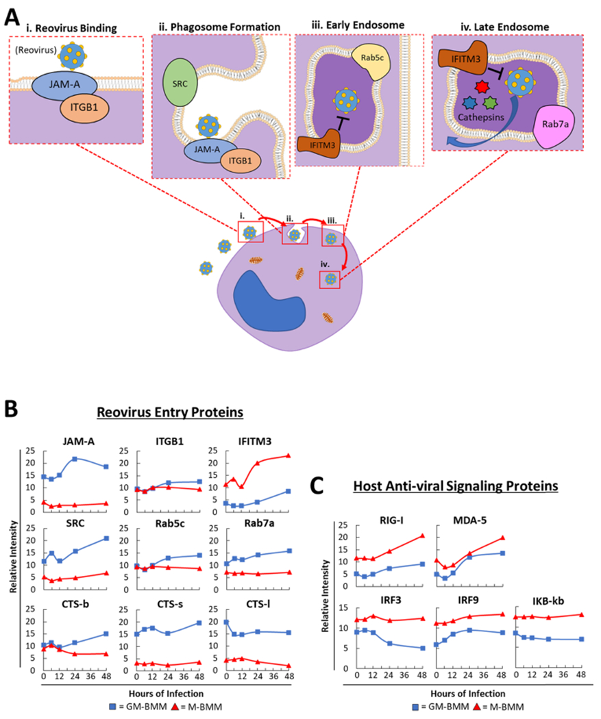
Figure 3.

Differential viral entry and antiviral proteins between GM-BMMs and M-BMMs. (A) (i-iv) Schematic representation of reovirus infecting a host cell. Cartoon representation depicts proteins involved in proper reovirus binding, phagosome formation, and early- and late-endosome composition. (B) Relative abundance of different proteins involved in reovirus binding [from (A)], endocytosis, and uncoating between GM-BMMs and M-BMMs. (C) Relative abundance of key host cell antiviral signaling proteins.
