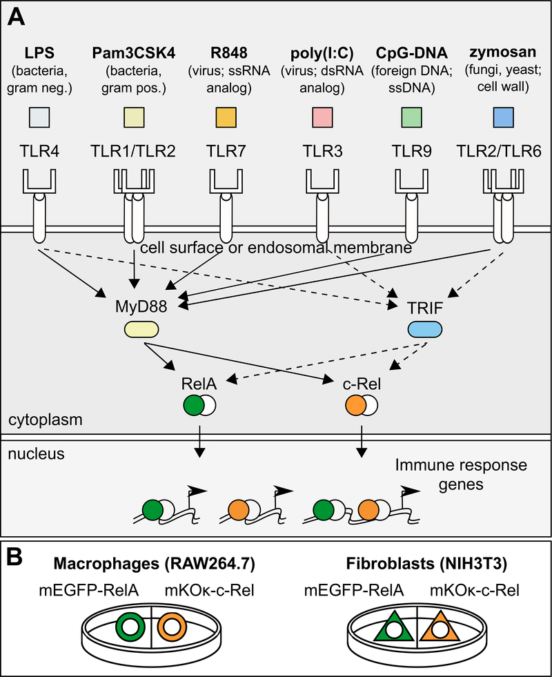
Fig. 1. Experimental design for imaging multiple NF-κB subunits in two cell types.
(A) Macrophages (RAW264.7 cells) and fibroblasts (NIH3T3 cells) were treated with an array of TLR ligands representing bacterial, viral, and fungal pathogens. Portrayed are the TLR ligands that produced detectable NF-κB activation in this study, as well as the targeted TLRs. LPS activates TLR4 (48); Pam3CSK4 activates TLR1/TLR2 heterodimers (49); R848 activates TLR7 (TLR7 and TLR8 in humans) (50, 51); zymosan activates TLR2/TLR6 heterodimers (52) and dectin-1 (53) (not depicted); poly(I:C) activates TLR3 (54); CpG-DNA activates TLR9 (55, 56); not shown: flagellin activates TLR5 (57) and profilin activates TLR11/TLR12 heterodimers (58, 59). (B) Macrophages or fibroblasts stably transfected with mEGFP-RelA or mKOκ-c-Rel were cultured in the same dish, treated with each of the TLR ligands, and then subjected to live-cell imaging. ssRNA, single-stranded RNA; dsRNA, double-stranded RNA; ssDNA, single-stranded DNA.

