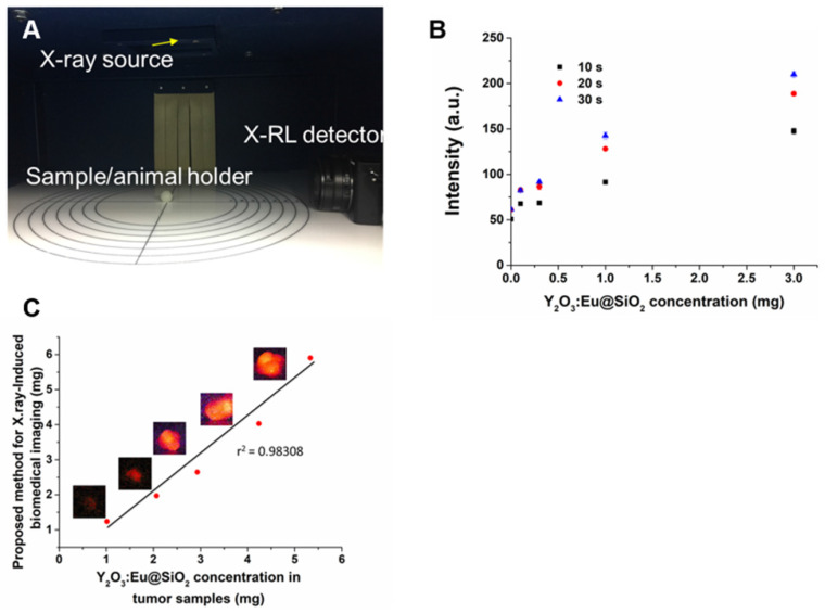
Figure 4.
X-RL image for deep tissue imaging activated by X-ray. (A) X-RL imaging system consists of a highly sensitive X-RL detector (CCD camera) and X-ray irradiator cabinet is enclosed within a light tight environment. (B) Exposure sensitivity of the X-RL imaging system was assessed by exciting Y2O3:Eu@SiO2 phantoms under 160 kVp X-rays. (C) The in-vivo calibration curve of X-RL intensity vs. particle concentration using subcutaneously inoculated SKOV3 tumor model.

