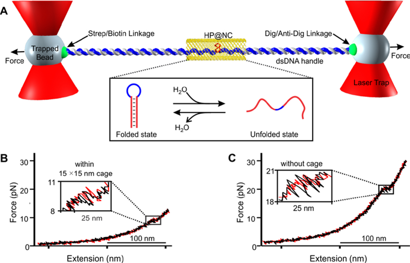
Figure 2.
Mechanical unfolding of the bcl-2 hairpin (HP) within DNA nanocage (NC). (A) The DNA construct in Figure 1 is tethered between two optically trapped beads via affinity interactions. Inset shows unfolding and refolding transitions of the hairpin inside nanocage (HP@NC). Strep, Dig and Anti-Dig represent streptavidin, digoxigenin and anti-digoxigenin respectively. Force versus extension curve of the DNA hairpin (B) within and (C) without 15×15 nm nanocage. Red and black traces indicate stretching and relaxing curves, respectively. Zigzag features in insets depict rapid unfolding/refolding transitions. Experiments were performed in a 20 mM Tris buffer (pH 7.8) supplemented with 100 mM KCl, 10 mM MgCl2, and 1 mM EDTA at 25 °C.

