
Figure 2.
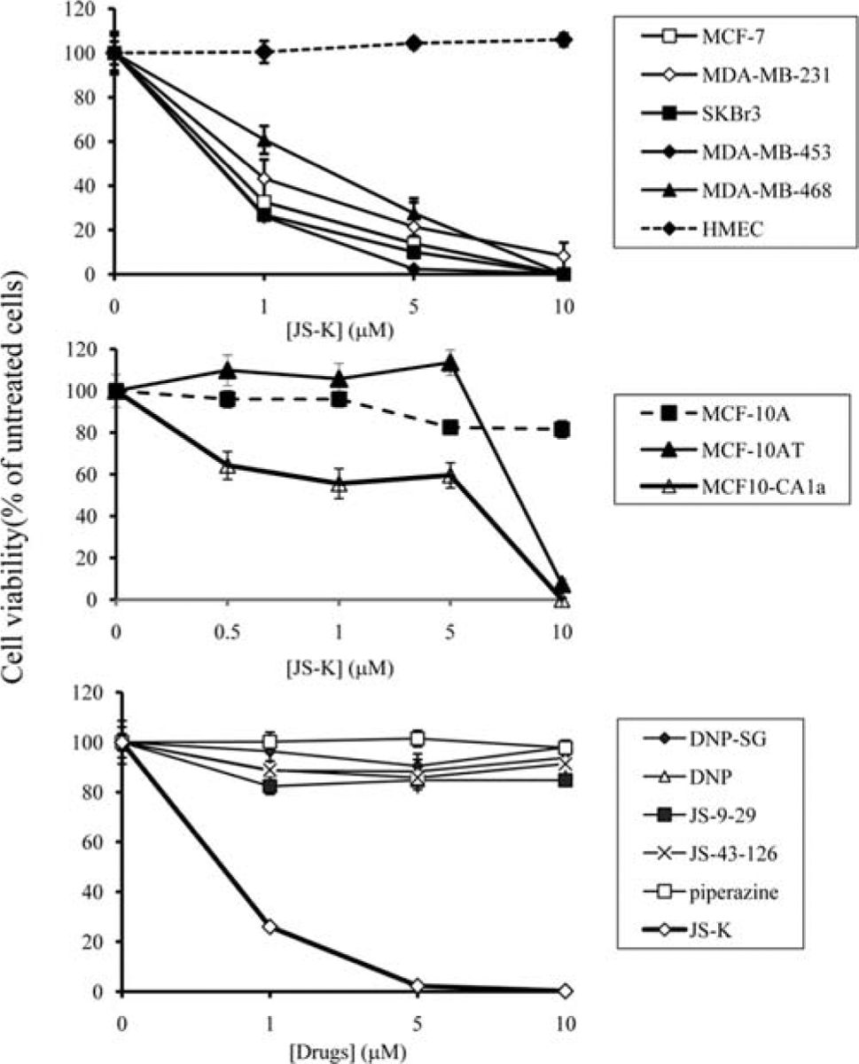
JS-K decreases the viability of breast cancer cells but not that of normal mammary epithelial cells. Breast cancer cells and normal HMECs were incubated with increasing concentrations (1, 5, 10 μM) of JS-K for 3 days (top panel). Cell viability was determined by the Promega Celltiter 96 AQueous non-radioactive proliferation assay. Data points represent the mean of five replicates. Data are expressed as mean percentage of untreated cells ± standard deviation. Non-transformed MCF-10A, premalignant MCF-10AT and fully malignant MCF-10CA1a mammary epithelial cells were incubated with increasing concentrations (1, 5, 10 μM) of JS-K for 3 days (middle panel). Cell viability was determined by the Promega Celltiter 96 AQueous non-radioactive proliferation assay. Data points represent the mean of five replicates. Data are expressed as mean percentage of untreated cells ± standard deviation. MDA-MB-453 breast cancer cells were treated with JS-K, its presumed metabolites (DNP-SG, DNP, JS-9-29, and piperazine), and a non-NO-releasing, non-arylating control (JS-43-126) for 3 days (bottom panel). Cell viability was determined by the Promega Celltiter 96 AQueous non-radioactive proliferation assay. Data points represent the mean of five replicates. Data are expressed as mean percentage of untreated cells ± standard deviation.