
Figure 3.
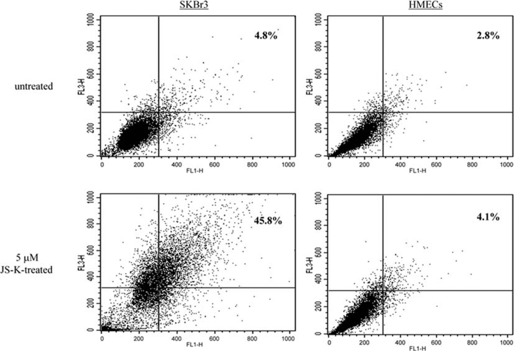
JS-K increases the formation of acidic vesicular organelles in breast cancer cells but not in normal mammary epithelial cells. SKBr-3 breast cancer cells and normal HMECs were incubated with JS-K (0, 5 μM) for 3 days, and stained with the acridine orange dye. FACS analysis was used to detect the green and the red fluorescence in the acridine orange-stained cells. FL1-H (x-axis) indicates green color intensity, while FL3-H (y-axis) shows red color intensity. Percentage of acridine orange-stained positive cells is the sum of percentage of cells in the upper left and the upper right quadrants.