
Figure 6.
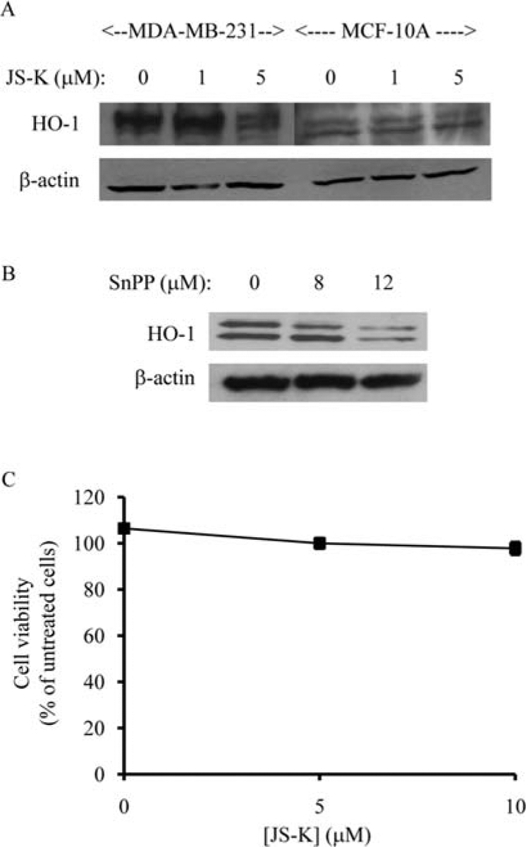
JS-K selectively decreases HO-1 expression in breast cancer cells. MDA-MB-231 breast cancer cells and MCF-10A mammary epithelial cells were treated with 0, 1, 5 μM of JS-K for 4 h (A). Membranes were incubated with anti-HO-1 antibody. ß-actin was used as a loading control. Protein bands were visualized by enhanced chemiluminescence. MCF-10A mammary epithelial cells were incubated with 0, 8, 12 μM of SnPP (HO-1 inhibitor) for 24 h (B). Membranes were incubated with anti-HO-1 antibody. ß-actin was used as a loading control. Protein bands were visualized by enhanced chemiluminescence. MCF-10A cells were preincubated with 12 μM of SnPP for 24 h before being treated with JS-K (0, 5, 10 μM) for 3 days (C). Cell viability was determined by the Promega Celltiter 96 AQueous non-radio-active proliferation assay. Data points represent the mean of five replicates. Data are expressed as the mean percentage of untreated cells ± standard deviation.